How Rehabilitative Ultrasound Imaging Can Transform Your Pelvic Health Practice
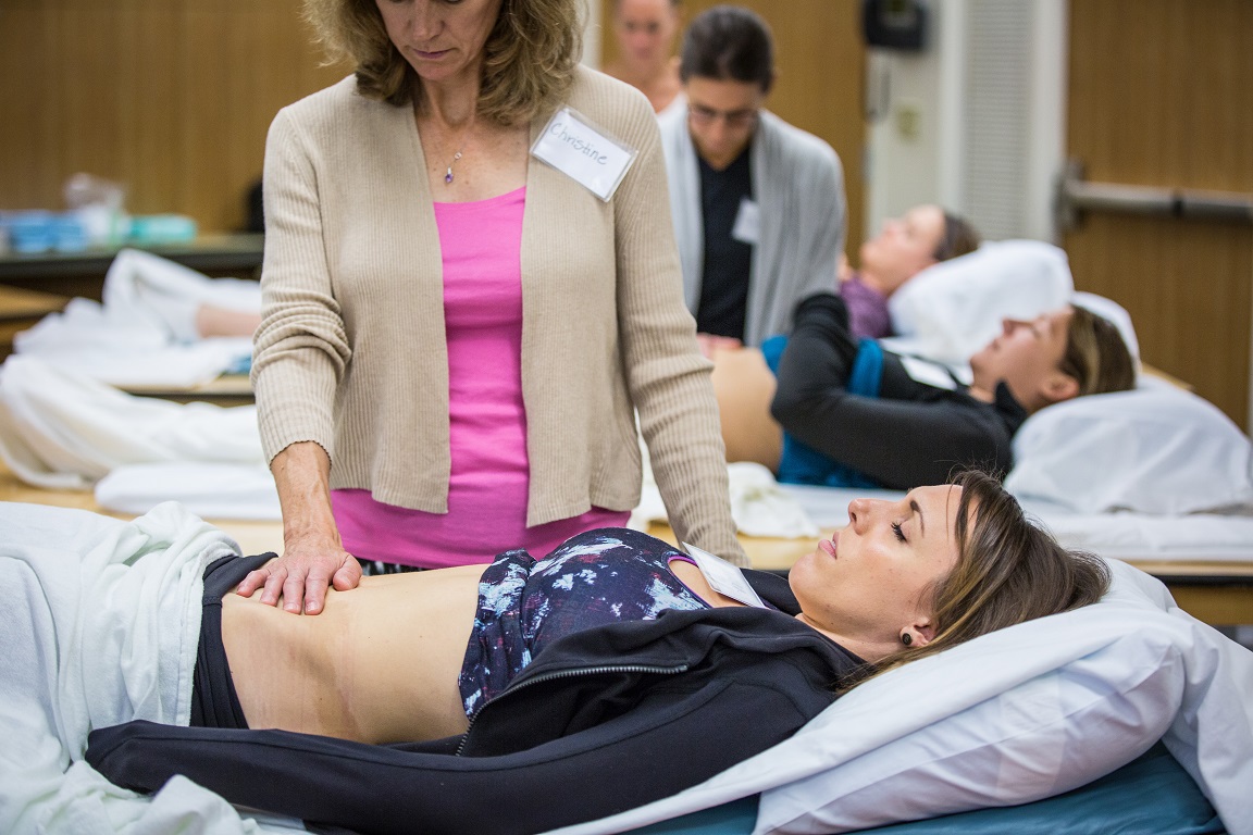
Rehabilitative ultrasound imaging is a clinical tool that can change the way you practice. I have often shared with other clinicians how much the use of ultrasound imaging has influenced how I approach patients with chronic back or sacroiliac joint pain. Using ultrasound imaging allows for a way to assess the deeper core muscles, which may be more difficult to palpate on some individuals. Being able to view the activation in these muscles can inform the therapist whether the patient is properly activating their core or relying on a less ideal strategy.
Seeing the Core Muscles That Are Hard to Reach
One of the most valuable things about rehabilitative ultrasound imaging in pelvic health is what it shows us about the deeper core muscles. These are muscles that conventional palpation simply cannot reach reliably in every patient. With ultrasound imaging, we can observe in real time whether a patient is using a proper activation strategy or compensating in a way that looks adequate on the surface but is not providing the stability they need.
That kind of information changes treatment. It gives both the clinician and the patient something concrete to work with, and it often unlocks progress that had stalled.
A Game Changer for Incontinence and Prolapse
The use of rehabilitative ultrasound imaging has also been a game changer in treating incontinence and prolapse patients. Not only does it enable me to view activation in the pelvic floor, but also the supportive function of the pelvic floor. For some patients, that supportive function is exactly what has been missing. Being able to show them what is happening in their own body, in real time, is often what finally moves treatment forward.
When Ultrasound Made All the Difference: A Patient Story
I recently began working with a patient who is a semi-professional athlete. She was 14 months postpartum and seeking care for prolapse symptoms and discomfort. This patient understood the importance of the pelvic floor and had sought out pelvic floor rehab immediately following delivery. She was approved to return to exercise and at the sixth minute of activity felt a prolapse occur.
After returning and continuing with pelvic health therapy, she still was not seeing progress with respect to her symptoms. There was real pressure mounting because she had qualified for an international level event in her sport that was six months away.
When I evaluated her, I identified that she was able to activate her pelvic floor while in a supine position, but not when standing or during a motor task. Using rehabilitative ultrasound imaging allowed her to visualize what it felt like to do a proper contraction while in standing. This was transformative. It helped her learn to engage her pelvic floor in a weight bearing position, which improved the supportive function of the pelvic floor and allowed her to begin engaging it during her sports activity.
It did take time and a lot of practice. But the addition of ultrasound imaging was what made the difference between her earlier attempts at pelvic rehab and this course of treatment.
About the Instructor: Allison Ariail, PT, DPT, CLT-LANA, BCB-PMD, PRPC
Allison Ariail, PT, DPT, CLT-LANA, BCB-PMD, PRPC is the creator of the Rehabilitative Ultrasound Imaging courses at Herman & Wallace and currently serves as Director of Education. A physical therapist since 1999, Allison holds a Doctor of Physical Therapy from Boston University. She is board certified by the Lymphology Association of North America (2011), board certified in Biofeedback Pelvic Muscle Dysfunction (2012), and earned her Pelvic Rehabilitation Practitioner Certification in 2014. She is a published researcher, a co-author in Healing in Urology, and a nationally recognized lecturer on ultrasound imaging, lymphedema, and pelvic floor dysfunction. Allison practices at Inspire Physical Therapy and Wellness in the Denver metro area, treating men, women, and children across a wide range of pelvic health conditions.
Join Us in Edmond, Oklahoma: April 17 to 19, 2026
This three-day course covers transabdominal viewing of the pelvic floor, abdominal wall, and spinal muscles as well as transperineal imaging that allows us to view the supportive function of the pelvic floor. Topics include:
- Real-time imaging of the transverse abdominals, rectus abdominis, deep multifidus, levator ani, bladder, bladder neck, urethra, and vagina
- Transabdominal and transperineal viewing methods
- Hands-on lab time with ultrasound machines provided by course sponsors
- Clinical application for lumbopelvic pain, pelvic organ prolapse, and urinary incontinence
You will love learning to use this clinical tool and seeing the changes it makes for your patients. Register for Rehabilitative Ultrasound Imaging: Pelvic Health and Orthopedic Topics in Edmond, OK at hermanwallace.com.
By accepting you will be accessing a service provided by a third-party external to https://www.hermanwallace.com/
All Upcoming Continuing Education Courses

Rehabilitative Ultrasound Imaging Pelvic Health Satellite Lab Course - Self-Hosted - April 17 - 19 2026
Apr 17 2026 - Apr 19 2026

Rehabilitative Ultrasound Imaging Pelvic Health Satellite Lab Course - Edmond OK - April 17 - 19 2026
Apr 17 2026 - Apr 19 2026

Pelvic Function Level 2B - Satellite - Bethpage NY - April 18 - 19 2026 - SOLD OUT
Apr 18 2026 - Apr 19 2026

Pelvic Function Level 1 - Satellite - Seattle WA - April 18 - 19 2026 - SOLD OUT
Apr 18 2026 - Apr 19 2026

Pelvic Function Level 1 - Satellite - Salt Lake City UT - April 18 - 19 2026 - SOLD OUT
Apr 18 2026 - Apr 19 2026
Pelvic Function Level 1 - Satellite - Newberg OR - April 18 - 19 2026 - SOLD OUT
Apr 18 2026 - Apr 19 2026

Menopause Transitions and Pelvic Rehab - Remote Course - April 18 - 19 2026
Apr 18 2026 - Apr 19 2026

Pelvic Function Level 1 - Satellite - Wichita KS - April 18 - 19 2026 - SOLD OUT
Apr 18 2026 - Apr 19 2026
Oncology of the Pelvic Floor Level 2A - Remote Course - April 18 - 19 2026
Apr 18 2026 - Apr 19 2026

Pelvic Function Level 1 - Satellite - St. Augustine FL - April 18 - 19 2026
Apr 18 2026 - Apr 19 2026

Pelvic Function Level 1 - Satellite - Minneapolis MN - April 18 - 19 2026 - SOLD OUT
Apr 18 2026 - Apr 19 2026


Pelvic Function Level 1 - Satellite - Colorado Springs CO - April 18 - 19 2026
Apr 18 2026 - Apr 19 2026


Pelvic Function Level 2B - Satellite - Nashotah WI - May 9 - 10 2026 - SOLD OUT
May 9 2026 - May 10 2026


Mobilization of the Myofascial Layer - Satellite Lab Course - Torrance CA - May 15 - 17 2026
May 15 2026 - May 17 2026
Mobilization of the Myofascial Layer - Satellite Lab Course Self-Hosted - May 15 - 17 2026
May 15 2026 - May 17 2026

Mobilization of the Myofascial Layer - Satellite Lab Course - McKinney TX - May 15 - 17 2026
May 15 2026 - May 17 2026
Mobilization of the Myofascial Layer - Satellite Lab Course - Bradenton FL - May 15 - 17 2026
May 15 2026 - May 17 2026

Mobilization of the Myofascial Layer - Satellite Lab Course - Atlanta GA - May 15 - 17 2026
May 15 2026 - May 17 2026

Pediatrics Level 2 - Adv Pediatric Bowel and Bladder Disorders - Remote Course - May 16 - 17 2026
May 16 2026 - May 17 2026


Pelvic Function Level 1 - In-Person - Bridgewater NJ - May 16 - 17 2026 - SOLD OUT
May 16 2026 - May 17 2026