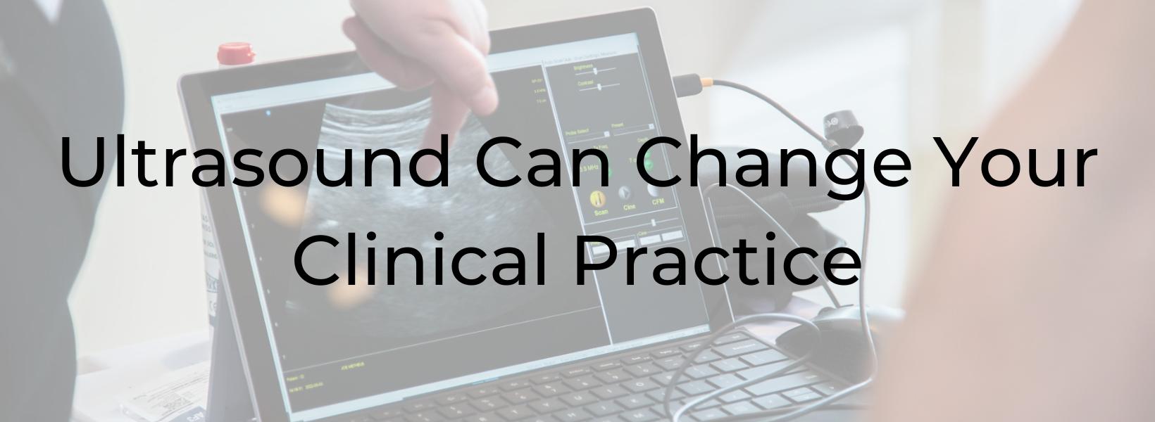
Ultrasound Can Change Your Clinical Practice
Allison Ariail, PT, DPT, CLT-LAANA, BCB-PMD is one of the creators of the Herman & Wallace Oncology of the Pelvic Floor Course Series as well as created the Rehabilitative Ultraosund courses.. Allison Ariail is a physical therapist who started working in oncology in 2007 when she became certified as a lymphatic therapist. You can join Allison in her upcoming two-day course September 29-30 (Rehabilitative Ultrasound: Orthodedic Topics) or the extended three-day course Rehabilitative Ultrasound: Women's Health and Orthodedic Topics that is through October 1st. You can also see Allison at HWConnect in the vendor hall where she will be doing ultrasound demonstrations. HWConnect is being held in Seattle, Washington this October 6-8, 2023.
“The widespread use of imaging has the potential to change the management of pelvic floor morbidity, such as urinary and anal incontinence, pelvic organ prolapse and related conditions ………. the insights provided by real-time imaging will enhance diagnostic and therapeutic capabilities.”1 This is a quote from an opinion article in Obstetrics and Gynecology by Hans Peter Dietz. Dietz has been researching the use of ultrasound and how it can assist in the diagnosis and treatment of pelvic floor disorders for years. I couldn’t agree more with this quote! Over the last 20 years that I have been using US imaging in my practice, I have seen more and more clinicians embrace ultrasound and let it change how they treat patients.
I have always told clinicians that using ultrasound will not only change your practice, but it will change patients’ lives. From using it to help postpartum women relearn to activate their core muscles, to helping chronic back pain patients that don’t get better with traditional treatment, to recent advances in a more targeted training method for postprostatectomy patients; using ultrasound will influence your treatments and help patients get back to living the lives and doing what they love to do.
In my experience, using ultrasound has been a rewarding experience to see how this tool can assist patients to meet their goals. There is something about the visual biofeedback and seeing what it happening inside the body to help patients have that “aha” moment where they get excited about treatment, become more motivated, and start to see results that they may not have had before. I recently had a patient that came to see me after having multiple sessions of therapy for urinary incontinence after a prostatectomy with another very qualified clinician. He was still having some leakage with sit-to-stand and during his tennis game. We were able to use ultrasound imaging to see that when he was performing a task that required more physical effort, he was no longer holding the pelvic floor contraction and was in fact using the incorrect strategy that increased pressure on his bladder. The visual biofeedback provided by ultrasound helped him learn to properly engage his pelvic floor with sit-to-stand and during athletic maneuvers. I only saw this patient for two treatments before I received a call from him telling me that he had seen so much progress and was doing so well that he felt he was ready to graduate from therapy.
Ultrasound can change your clinical practice. You can use it with back and sacroiliac joint patients, urinary and fecal incontinence patients, prolapse patients, post-prostatectomy patients, bowel patients, postpartum patients,….the list goes on! The prices of ultrasound units continue to go down and make it much more affordable for clinicians to be able to afford. I would recommend considering adding this tool to your list of available tools in your clinic.
At HWConnect, you will hear speakers discuss using ultrasound in their research and can see a demo of ultrasound use for a pelvic floor patient. You can also learn to use ultrasound in your practice with a Rehabilitative Ultrasound Imaging course offered through Herman and Wallace this September, as well as several dates for next year! If you do decide to get this exciting new tool, I promise you won’t be disappointed once you learn to use it and implement it into your clinical practice!
References:
- Dietz, HP. Ultrasound in the investigation of pelvic floor disorders. Curr Opin Obstet Gynecol. 2020; Dec; 32(6):431-440.

Rehabilitative Ultrasound Imaging - Women's Health and Orthopedic Topics - September 29 - October 1, 2023
*3 Day course (Friday-Saturday-Sunday) with internal and external labs
Experience Level: Intermediate
Contact Hours: 22.5
Description: This three-day course is designed to provide instruction in the generation and interpretation of rehabilitative ultrasound images as they relate to the pelvic girdle. Both lecture and lab material will be presented that will allow the participant to immediately incorporate the use of rehabilitative ultrasound imaging into their evaluation and treatment plans for patients with diagnoses including lumbopelvic pain and instability, pelvic organ prolapse, and urinary incontinence.
The course comprises of lectures and labs that present both ideal and abnormal responses of real-time imaging of the transverse abdominals, rectus abdominis, deep multifidus, levator ani, bladder, bladder neck, urethra, and vagina during contraction and Valsalva. Imaging methods used during labs will consist of transabdominal viewing as well as transperineal/translabial viewing methods. Prior experience with perineal and vaginal assessments is required to take the course. During labs, participants will be able to practice using machines provided by course sponsors.
This is an intermediate-level rehabilitative ultrasound imaging course intended for therapists having sound knowledge of training techniques for the local stabilizing muscles of the pelvic floor, transverse abdominals, and deep multifidi muscles and experience with internal vaginal exams. Prior coursework regarding the local stabilizing muscles (Diane Lee/ Linda Joy Lee, Paul Hodges, Pelvic Floor/Pelvic Girdle) is required.
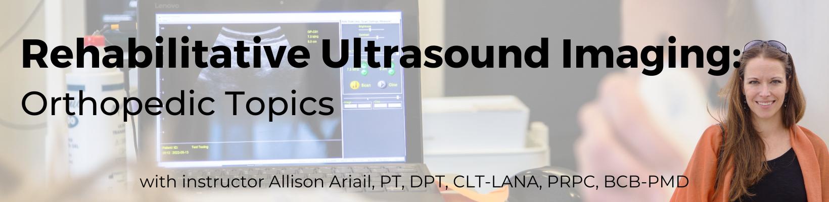
Rehabilitative Ultrasound Imaging - Orthopedic Topics - September 29-30, 2023
*2 Day course (Friday-Saturday) with external labs only
Price: $475
Experience Level: Intermediate
Contact Hours: 14
Description: This two-day course is designed to provide instruction in the generation and interpretation of rehabilitative ultrasound images as they relate to the pelvic girdle and low back stabilization. Both lecture and lab material will be presented that will allow the participant to immediately incorporate the use of rehabilitative ultrasound imaging into their evaluation and treatment plans for patients with diagnoses including lumbo-pelvic pain and instability.
This course comprises of lectures and labs that present both ideal and abnormal responses of real-time imaging of the transverse abdominals, rectus abdominis, deep multifidus, levator ani, bladder, and bladder neck. During labs, participants will be able to practice using machines provided by course sponsors.
This is an intermediate level rehabilitative ultrasound imaging course intended for therapists having a sound knowledge in training techniques for the local stabilizing muscles of the pelvic floor, transverse abdominals, and deep multifidi muscles. Prior coursework regarding the local stabilizing muscles is required.
By accepting you will be accessing a service provided by a third-party external to https://www.hermanwallace.com/
All Upcoming Continuing Education Courses

Pelvic Function Level 1 - Satellite - Milwaukee WI - July 12 - 13 2025 - SOLD OUT
Jul 12 2025 - Jul 13 2025

Modalities and Pelvic Function - In-Person - Houston TX - July 12 - 13 2025
Jul 12 2025 - Jul 13 2025

Pelvic Function Level 1 - Satellite - Denver CO - July 12 - 13 2025 - SOLD OUT
Jul 12 2025 - Jul 13 2025


Pelvic Function Level 1 - Satellite - Des Moines IA - July 26 - 27 2025 - SOLD OUT
Jul 26 2025 - Jul 27 2025

Pelvic Function Level 1 - Satellite - Minneapolis MN - July 26 - 27 2025 - SOLD OUT
Jul 26 2025 - Jul 27 2025

Pelvic Function Level 1 - Satellite - Paso Robles CA - July 26 - 27 2025 - SOLD OUT
Jul 26 2025 - Jul 27 2025

Pelvic Function Level 1 - Satellite - St. Augustine FL - July 26 - 27 2025
Jul 26 2025 - Jul 27 2025

Pelvic Function Level 1 - Satellite - Virginia Beach VA - July 26 - 27 2025 - SOLD OUT
Jul 26 2025 - Jul 27 2025


Pelvic Function Level 1 - In-Person - Chicago IL - August 2 - 3 2025 - SOLD OUT
Aug 2 2025 - Aug 3 2025


Menopause Transitions and Pelvic Rehab - Remote Course - August 9 - 10 2025
Aug 9 2025 - Aug 10 2025


Pelvic Function Level 2C - Satellite - Palm Beach FL - August 16 - 17 2025
Aug 16 2025 - Aug 17 2025

Pelvic Function Level 2C - Satellite - New Orleans LA - August 16 - 17 2025
Aug 16 2025 - Aug 17 2025
Pelvic Function Level 2C - Satellite - Paso Robles CA - August 16 - 17 2025
Aug 16 2025 - Aug 17 2025


Pediatrics Level 1 - Treatment of Bowel and Bladder Disorders - Remote Course - August 23 - 24 2025
Aug 23 2025 - Aug 24 2025

Pelvic Function Level 1 - Satellite - Jacksonville FL - August 23 - 24 2025
Aug 23 2025 - Aug 24 2025

Pelvic Function Level 1 - Satellite - Chicago IL - August 23 - 24 2025 - SOLD OUT
Aug 23 2025 - Aug 24 2025


Pelvic Function Level 1 - Satellite - Hermosa Beach CA - August 23 - 24 2025
Aug 23 2025 - Aug 24 2025

Rehabilitative Ultrasound Imaging Pelvic Health Satellite Lab Course - Self-Hosted - September 5 - 7 2025
Sep 5 2025 - Sep 7 2025
Rehabilitative Ultrasound: Orthopedic Topics Satellite Lab Course - Self-Hosted - September 5 - 6 2025
Sep 5 2025 - Sep 6 2025

Rehabilitative Ultrasound Imaging Pelvic Health Satellite Lab Course - Indianapolis IN - September 5 - 7 2025
Sep 5 2025 - Sep 7 2025
