

H&W is proud to be able to present a new remote course on ethics from new faculty member, and Sr. TA, Mora Pluchino, PT, DPT, PRPC. Mora is a graduate of Stockton University with a BS in Biology (2007) and Doctorate of Physical Therapy (2009). She has been working at Bacharach Institute for Rehabilitation ever since graduation and has experience in a variety of areas and settings, working with children and adults, including orthopedics, bracing, neuromuscular issues, vestibular issues, and robotics training.
Mora began treating Pelvic Health patients in 2016 and has experience treating women, men, and children with a variety of Pelvic Health dysfunction. In 2020, she opened her own "after hours" virtual practice called Practically Perfect Physical Therapy Consulting to help meet the needs of more clients and has been a guest lecturer for Rutgers University Blackwood Campus and Stockton University for their Pediatric and Pelvic Floor modules and has been a TA with Herman & Wallace since 2020 with over 150 hours of lab instruction experience. Mora authored and instructs Ethical Concerns for Pelvic Health Professionals.
● Your employer expects you to be responsible for supervising and billing for three orthopedic clients while you are in a private treatment area with a pelvic health patient. They say that you’re fine because the aide will be assisting the patients with their program.
● You are a new graduate, and your employer sends you to Pelvic Floor Level 1 and expects you to start up a Pelvic Health Program at the facility. Your first scheduled patient is a diagnosis you didn’t learn about and don’t feel comfortable treating, but your student loan payments have started, and you need this job.
● The word has gotten out and referrals are flowing into your clinic after hearing what a life-changing service your Pelvic Health program provides. You have a 6 month long waiting list and your front desk person asks you how to organize the list. Options include prioritizing based on the severity of issues, first come/ first serve, based on past history with the facility, etc.
Put yourself in these situations and reflect. How would you feel? What would you do? All of the situations above have a common theme. They present Ethical Concerns for Pelvic Health Professionals. Ethical situations are very common in day-to-day practice for any health care professional. Treating in the pelvic region can add additional complications due to the level of intimacy and vulnerability in this care.
Ethics by definition is “the division of philosophy concerned with how a person should behave in a matter that is considered morally correct or good” that gives us standards, virtues, and rules (Boone, 2017). The professional code of ethics for each professional category and is grounded in virtue ethics. Virtue ethics includes four main concepts including balance of harms and benefits, doing no harm, justice, and autonomy (Kirsch, 2005)
Whether a practitioner is a physical therapist/ physical therapist assistant, occupational therapist, psychologist, social worker, nurse, doctor, or physician’s assistant, their governing professional organization will have a code of ethics and conduct to help guide these practitioners in good decision making. There are many ethical decision-making models to help guide an individual through the process of identifying, defining, examining, and then problem solving any ethical occurrence.
The RIPS Model, originally presented by Arlanian, Swisher & Davis in 2005, presents an ethical framework to help guide practitioners through the steps needed to address ethical concerns. The world of ethical decision-making is typically one of many options, typically being more “gray area” than simply “black” and “white.” Ethical Concerns for Pelvic Health Professionals is designed to help make these decisions easier to define, examine, discuss, and address so we can have these tough conversations!
Ethical Concerns for Pelvic Health Professionals is a one-day remote course that covers ethical considerations for professionals working in the area of Pelvic Health. In general, Health Care Professionals have many day-to-day ethical considerations to “do no harm.” This includes basic decisions for billing, patient care, safety, and compliance. The purpose of this class is to explore the ethical challenges Pelvic Health practitioners may experience including consent, managing trauma and abuse, and preventing misconduct. To learn more join us in Ethical Concerns for Pelvic Health Professionals on June 18, 2022!
Research:
Boone, B. (2017). Ethics 101: From altruism and utilitarianism to bioethics and political ethics, an exploration of the concepts of right and wrong. Adams Media.
Kirsch, N. (2005). Ethics in Physical Therapy: A Case-Based Approach. McGraw Hill Publications. American Physical Therapy Association.
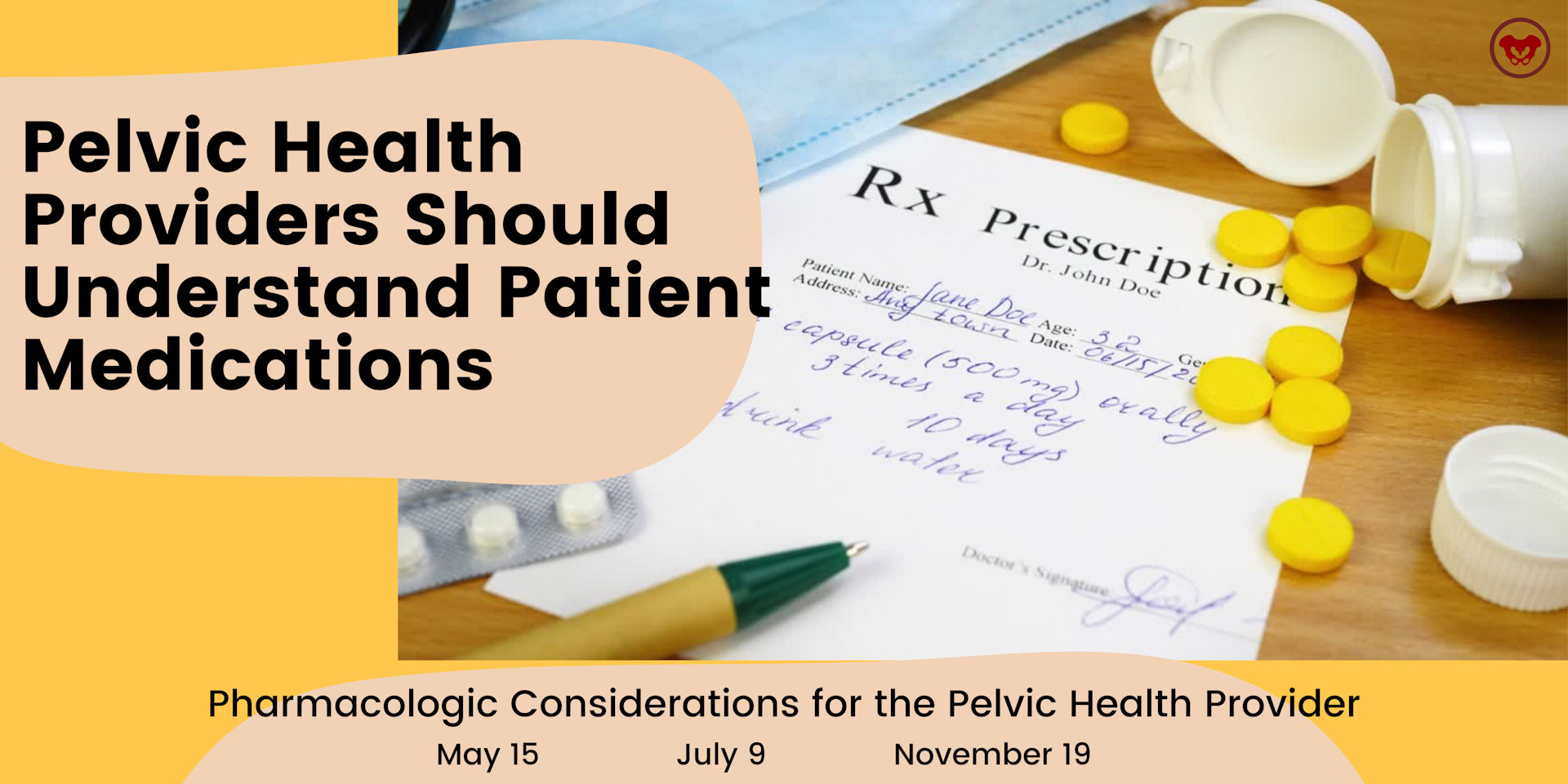

Kristina Koch, PT, DPT, is a board-certified women’s health physical therapist and certified lymphedema therapist who is the author/instructor of the remote course Pharmacologic Considerations for the Pelvic Health Provider. She has been treating pelvic health dysfunction in all ages and genders since 2001. Kristina works in private practice in Colorado Springs, CO, and has served as a guest lecturer for the pelvic health content at Regis University in Denver and the University of Colorado.
Did you know that the median length of time a primary care provider spends with a patient is 15 minutes? This breaks down to approximately 5 minutes for the patient to talk about their health concerns and 5 minutes for the health care provider to speak, with the rest being applied toward paperwork (1). After this appointment, the patient may not be seen again for several months or even a year. Medication side effects that impact the patients’ quality of life, or create new symptoms, can be easily overlooked.
I created the course, Pharmacologic Considerations for the Pelvic Health Provider because, with direct access to physical and occupational therapy services, a greater responsibility has been placed upon the therapist to take on more of a role as a primary care provider. We must ensure that all factors contributing to a patient’s signs and symptoms are considered. This includes reviewing and assessing if medications or supplements may be causative.
As therapists, we typically spend more time with our patients than their primary care providers. We are seeing them for longer periods during a treatment session and will see them numerous times throughout a month or longer. As a result, our patients have a greater opportunity to discuss their signs and symptoms with us versus their physician or primary care provider. Therefore, understanding how medications may be impacting a patient is essential. Being able to educate the patient about the side effects of the medications they are taking and how the medications may be contributing to their complaints can enhance the value of the treatment session.
By possessing this knowledge and understanding of the medications that are prescribed to treat pelvic health, therapists can have educated conversations with our patients and other health care providers involved in their care. The ability to discuss the most recent medications and supplements or complementary alternatives, that may minimize side effects or have fewer impacts on quality of life and enhance function, is an integral part of patient care.
Join Kristina to dive into Pharmacologic Considerations for the Pelvic Health Provider. This one-day, virtual course will discuss the importance of understanding pharmacology and review commonly prescribed medications. Kristina also spends time explaining current research pertaining to the pharmacologic treatment of numerous pelvic and reproductive health conditions, medication side effects, drug interactions, and non-pharmacologic alternatives. Medications discussed include those for constipation and GI dysfunction, pelvic pain conditions (including vulvodynia, chronic prostatitis, and endometriosis), as well as medications and side effects in Gender-Affirming Care for patients who are transitioning.
Upcoming 2022 course dates for Pharmacologic Considerations for the Pelvic Health Provider include:
References:
- Tai-Seale, M., McGuire, T. G., & Zhang, W. (2007). Time allocation in primary care office visits. Health Services Research. 42(5), 1871-1894. Doi:10.1111/j.175-6773.2006.00689.x
- Ciccone, C. D. (2007). Pharmacology in Rehabilitation. (4th). F.A. Davis Company.
This week the interviewer becomes the interviewee as Stacey Futterman Tauriello sits down to interview Holly Tanner on the Male Pelvic Floor course.
Stacey Futterman reached out to Holly Tanner back in 2007 through a phone call to see if they could partner on a lecture covering male pelvic pain. The two had never met in person but decided to collaborate on the three-hour APTA National Conference lecture. Holly shares "I still recall the frequent glances I made to match the person behind the voice I had heard for so many long phone calls.”
This presentation was developed into a two-day education course for Herman & Wallace and contained lectures on male anatomy, post-prostatectomy urinary incontinence, pelvic pain, and sexual health and dysfunction. The big question of the time was “should we allow men to attend?” Holly puts this in perspective, “As strange as this question now seems, it speaks volumes about the world of pelvic health at that time; mostly female instructors taught mostly female participants about mostly female conditions.”
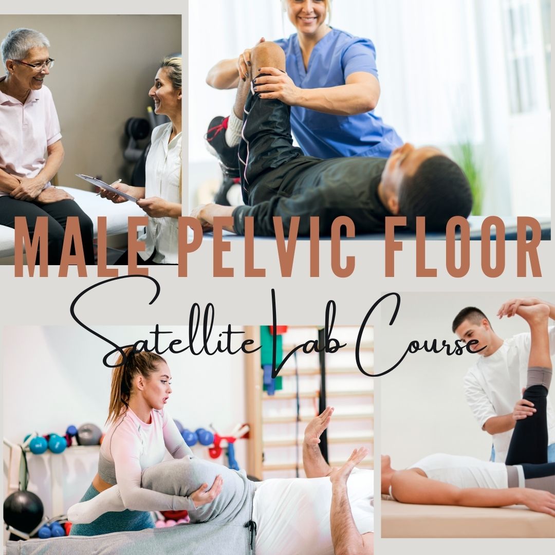
The Male Pelvic Floor course was first taught in 2008 and has since been expanded to include 22 contact hours. This current content includes 7 pre-recorded lectures and 2 full days of live lectures and labs, allowing more time for hands-on skills in examination and treatment. The schedule still covers bladder, prostate, sexual health, and pelvic pain, and further discusses special topics like post-vasectomy syndrome, circumcision, and Peyronie’s disease.
Because the course often has providers in attendance who have not completed prior pelvic health training, instruction in basic techniques is included. For the experienced therapists, there are multiple lab “tracks” that offer intermediate to advanced skills that can be practiced in addition to the basic skills. Holly adds, “One of the more valuable conversations that we have in the course is how to create comfort and ease in when for most us, we were raised in a culture (and medical training) where palpation of the pelvis was not made comfortable. Hearing from the male participants about their bodies, how they are affected by cultural expectations, adds significant value as well.”
In 2017 Herman & Wallace faculty member, Heather Radar submitted a blog where she wrote about a note that was left on her doorstep by the wife of an older gentleman who had chronic male pelvic pain. When looking into writing this blog I kept coming back to this past blog by Heather, and I have decided to share an abridged version today to accompany Holly Tanner's short interview discussing the Male Pelvic Floor Satellite Lab Course.
Recently, a note was left at my doorstep by the wife of an older gentleman who had chronic male pelvic pain. His pain was so severe, that he could not sit, and he lay in the back seat of their idling car as his wife, having exhausted all other medical channels available to her, walked this note up to the home of a rumored pelvic floor physical therapist who also treated men. The note opened with how she had heard of me. She then asked me to contact her about her husband’s medical problem. It ended with three words that have vexed me ever since…we are desperate. We Are Desperate.
Unlike so many men with chronic pelvic pain, he had at least been given a diagnostic cause of his pain, pelvic floor muscle dysfunction, rather than vaguely being told it was just a prostate issue. However, the therapists that had been recommended by his doctor only treated female pelvic dysfunction.
My first thought after reading the note was, “I bet shoulder or knee therapists don’t get notes like this on their doorstep.” My next thought, complete with facepalm, “THIS HAS TO STOP! Pelvic floor rehab has got to become more accessible”.
Pelvic floor therapists see all people including men, women, and transgender. They treat the pediatric, adult, and geriatric populations. They treat pelvic floor disorders in the outpatient, home health, and SNF settings. They treat elite athletes and those with multiple co-morbidities using walkers. They can develop preventative pelvic wellness programs and teach caregivers how to better manage their loved one’s incontinence. This is due to one simple fact: No matter the age, gender, level of health, or practice setting, every patient has a pelvic floor.
The pelvic floor should not be regarded as some rare zebra in clinical practice when it is the workhorse upon which so many health conditions ride. It interacts with the spine, the hip, the diaphragm, and vital organs. It is composed of skin, nerves, muscles, tendons, bones, ligaments, lymph glands, and vessels. It is as complex and as vital to function and health as the shoulder or knee is, and yet students are lucky if they get a “pelvic floor day” in their PT or OT school coursework.
I call for every therapist, specialist, and educator to learn more about the pelvic floor. If you only treat pelvic dysfunction in women, please consider expanding your specialty to include men. The guys really need your help. You literally may be the only practitioner around that has the skills to treat these types of problems. Yes, the concerns you have about privacy and feeling comfortable are valid. But, you are not alone in this. Smart people like Holly Tanner have figured all that stuff out for you and can guide you on how to expertly treat in the men’s health arena.
The Silver Lining. Thanks to the champions of pelvic floor rehab education, we’ve come a long way. The good news in this story is that this man’s doctor recognized early that he had pelvic floor muscle dysfunction and recommended that he see a pelvic floor physical therapist. The bad news-it took 2 years before he could find one. The ball is in our court, therapists. Let’s do better. Until there are no more men in the back seat, we still need to #LearnMoreAboutThePelvicFloor.
We need to continue to create more coursework and more clinical training opportunities so that the representation of those treating male patients improves. If you feel ready to take your training to the next level in caring for male pelvic dysfunction, join us for an upcoming Male Pelvic Floor Satellite Lab Course.

Male Pelvic Floor Satellite Lab Course is scheduled on several dates and satellite locations for 2022, including self-hosted course options. Dates include:
- April 23-24
- June 12-13
- September 24-25
- October 22-23
- December 3-4


Blog by Deanna Vaughn, PT, DPT who practices at Core and Pelvic Physical Therapy Clinic in Conway, Arkansas, this article was originally located at https://whatsupdownthere.info/colorectal-cancer-the-gut-and-the-butt/.
Colorectal cancer refers to cancerous cells within the colon or rectum. Need a quick anatomy review? Keep reading then!
The colon is another name for the large intestine, which is the long tube (nearly 5 FEET!) surrounding the small intestines (that snaky, jumbled tube in the middle of our bodies, which you can see below in the picture). It’s comprised of segments: the cecum (the little pouch that joins the small intestine to the large intestine) in the right lower abdomen, the ascending colon starting at the right lower part of your abdomen (coming off the cecum), and up to about the right side of your ribcage; the transverse colon that loops underneath the stomach and ribcage from right to left; the descending colon that extends down from the left side of your ribcage to the lower part of your left abdomen; and then the sigmoid colon that loops (in an s-shape) along the lower abdomen to the center of the body. At the end of the colon is the rectum, which pretty much connects the colon to the actual anus/anal opening for wastes to leave the body.
That being said, colorectal cancer can affect any part or segment of the colon and the rectum. If you have a family history of colorectal cancer, or if you have an inflammatory bowel disease (like Crohn’s disease or ulcerative colitis), then you may be at a higher risk for colorectal cancer. Other risk factors are the same for virtually any other health condition – genetics, no regular physical activity, poor diet, tobacco use, high alcohol consumption, etc.
So how would we know if it’s colorectal cancer – or precancerous cells, and how do we decrease our risk?
That’s where screening comes into play! Just like how someone may see their gynecologist annually and undergo the PAP smear every 1-3 years to check for any gynecological cancer (like cervical or labial cancer), someone may see their colorectal or gastrointestinal (GI) provider to check for colorectal cancer or disorders. Regular screening takes place around age 45 (although a person may be screened earlier if they are at higher risk or had a previous history of cancer).
What does screening look like?
There are a few tests that screen for colorectal cancer. These tests include stool tests, flexible sigmoidoscopy, and colonoscopy.
Stool tests – This pretty much involves you taking a sample of your stool via test kit provided to you, and returning it to your doctor/lab, where your stool is checked for any blood or other abnormal findings.
Flexible sigmoidoscopy – A thin, short tube with a light is inserted into the rectum. This allows your doctor to see any polyps or cancer within the rectum and lower part of the colon.
Colonoscopy – This is like the sigmoidoscopy, but with a longer tube. The longer tube allows your doctor to check for polyps/cancer inside the rectum and the entire length of the colon. Your doctor can also remove some polyps during this procedure if indicated.
Most people without any symptoms, abnormal findings or outstanding personal or family history of colorectal cancer will have these screening tests performed anywhere from 5-10 years.
What are the symptoms?
This is not an exhaustive list, but some symptoms may include:
- Bleeding, pain, and/or discomfort within the rectum/anus
- Blood in stool
- Abdominal pain and bloating
- Nausea/vomiting
- Difficulty or incomplete bowel evacuation
- Hemorrhoids
- Altered bowel habits (such as sudden constipation, diarrhea, change in stool consistency)
Now what are our treatment options?
Besides preventative measures – such as getting regular physical activity, improving our diet, etc., treatment looks similar to any other cancer treatment. This may look like chemotherapy, radiation therapy, immunotherapy, and/or surgery. Surgery may be indicated to remove polyps/tumors, or parts of the colon or rectum to eliminate cancerous growths. Thankfully though, regular screening of the colorectal region can find precancerous/cancerous cells early. Oftentimes, such as during a colonoscopy, your colorectal provider may go ahead and remove polyps that are abnormal or deemed precancerous at that time!
Now what about pelvic physical therapy? Can it possibly help?
Well, this is another condition (like Pelvic Congestion Syndrome in the previous blog post), where pelvic physical therapy is not the initial go-to or main treatment option. Individuals with colorectal cancer vary in several ways depending on staging/severity and overall health. Once again, pelvic therapy is a nice resource to utilize if you’re needing or wanting ways to manage your bowel symptoms.
Ways that pelvic PT CAN help may include: Teaching appropriate toileting – positioning to straighten out the anorectal angle and allow stool to pass more easily from the rectum; mechanics, such as exhaling smoothly when pushing for a bowel movement to prevent straining; Improving pelvic floor muscle function (strength, endurance, coordination) so that your body can delay defecation as needed and calm down bowel urges; and overall promoting health bowel habits by supporting your nutrition and keeping bowel movements regular.
Whether or not you (or someone you know) have colorectal cancer, developing healthy and safe bowel habits is key to a better quality of life. Working with your doctor and/or your team of providers is important in making sure your needs are addressed, but feel free to reach out to your local pelvic PT if you want more resources or guidance – even things like, “So, how SHOULD I be pooping??”
References & Resources
Brenner H, Chen C. The colorectal cancer epidemic: challenges and opportunities for primary, secondary and tertiary prevention. Br J Cancer. 2018;119(7):785-792. doi:10.1038/s41416-018-0264-x
https://www.cancer.org/cancer/colon-rectal-cancer.html
https://my.clevelandclinic.org/health/diseases/14501-colorectal-colon-cancer
Kuipers EJ, Grady WM, Lieberman D, et al. Colorectal cancer. Nat Rev Dis Primers. 2015;1:15065. Published 2015 Nov 5. doi:10.1038/nrdp.2015.65
Leslie A, Steele RJC. Management of colorectal cancerPostgraduate Medical Journal 2002;78:473-478. http://dx.doi.org/10.1136/pmj.78.922.473
Mármol I, Sánchez-de-Diego C, Pradilla Dieste A, Cerrada E, Rodriguez Yoldi MJ. Colorectal Carcinoma: A General Overview and Future Perspectives in Colorectal Cancer. Int J Mol Sci. 2017;18(1):197. Published 2017 Jan 19. doi:10.3390/ijms18010197
You YN, Lee LD, Deschner BW, Shibata D. Colorectal Cancer in the Adolescent and Young Adult Population. JCO Oncol Pract. 2020;16(1):19-27. doi:10.1200/JOP.19.00153


Dawn Sandalcidi PT, RCMT, BCB-PMD is known as the go-to expert in the field of pediatric pelvic health. She has been practicing for 40 years this May and has concentrated on the pediatric pelvic floor for 29 of those. When it comes to pediatric pelvic floor issues, there is so much more than bedwetting, and often the practitioner needs to look beyond the pelvic floor.
Despite the growing number of pelvic rehab specialists treating men and women with PF dysfunction, children in this patient population remain woefully under-served. This can cause undue stress for the child and family, as well as the development of internalizing and externalizing psychological behaviors. Many of the techniques used in pediatric pelvic therapy can be translated to the adult population. The question is ‘who’s the driver?’ In pediatrics, it is typically a bowel issue.
The Standard American Diet involves food that is high in calories, saturated fats, trans fats, added sugars, and sodium. It is also lacking in the intake of essential nutrients for the body like fiber, calcium, potassium, and vitamin D. This lack of dietary fiber can cause issues with the digestive tract as well as the colon leading to constipation. Bowel dysfunction including constipation can contribute to urinary leakage and urgency (1). Constipation accounts for approximately 5% of visits to pediatric clinics (2) proving that there is a need for practitioners to know how to treat these pediatric issues.
Dawn focuses much of her pediatric knowledge on her two courses: Pediatric Incontinence and Pelvic Floor Dysfunction (PEDs) and Pediatric Gastrointestinal Disorders (PEDsG). Pediatric pelvic floor basics are covered in PEDs, including instruction in anatomy, physiology, development of normal voiding reflexes and urinary control, and learning how to talk with child patients. Biofeedback and ultrasound (which Dawn fondly calls jelly belly) are also covered and can be helpful as less invasive procedures for children.
PEDsG goes beyond the pelvic floor and opens up the door to look at the big picture of the whole child. Dawn shares that almost 80% of her kiddos with chronic constipation present with diastasis rectus abdominus. They can also have hyperextension in the thoracic spine, and the rib cage is postally rotated – where the kids don’t know how to bring it down.
Dawn is also on the threshold of writing a pediatric pelvic pain course that she expects to be ready later this year. Pediatric pelvic pain is becoming more prevalent, and it can’t be treated the same way as in adults. Dawn explains that “children don’t understand, so we’re actually creating a pediatric pain neuroscience protocol. It is a bio-psycho-social approach, and we use fun things.”
Research tells us that 15% of kids per year will outgrow bedwetting. Children who suffer from bedwetting can feel ashamed and embarrassed, have self-esteem issues, or even act out. There are 5 basics of where you start with a pediatric patient that are taught in PEDs. Dawn also shares 5 basics in her e-book, BEDWETTING BOOTCAMP(3):
- Avoid things that can irritate the bladder
- Avoid drinking anything just before bed
- Drink throughout the day
- Empty your bladder throughout the day
- Avoid constipation and straining
Everything in Pediatric Incontinence and Pelvic Floor Dysfunction builds into Pediatric Gastrointestinal Disorders, and everything in PEDsG builds into Pediatric Pelvic Pain. The more practitioners who learn about the pediatric pelvic floor means that more kids get treated and the fewer adults that will have pelvic floor dysfunction. To learn more about treating pediatric pelvic health register for one of Dawn Sandalcidi’s upcoming courses:
Pediatric Incontinence and Pelvic Floor Dysfunction: August 27-28th
Pediatric Gastrointestinal Disorders: May 14-15, November 12-13
References:
- Sandalcidi (Spring 2018). Paediatric incontinence and pelvic floor dysfunction. Journal of Pelvic, Obstetric and Gynaecological Physiotherapy, 122, 5–8.
- Thibodeau B. A., Metcalfe P., Koop P. & Moore K. (2013) Urinary incontinence and quality of life in children. Journal of Pediatric Urology 9 (1), 78–83.
- Dawn Sandalcidi PT, RCMT, BCB-PMD. BEDWETTING BOOTCAMP Frustrated by Bed Wetting? The 5 Ways to Speed Starting Up Dry Tonight! www.kidsbowelbladderPMD.com. https://cdn.fs.teachablecdn.com/YfP5rUfiRtOJxXZSZUso


This is the second installment in our 3 part pediatric blog series written by Amanda Moe DPT, PRPC treats women, men, and children with disorders of the pelvis and pelvic girdleAmanda enjoys assistant teaching with the Herman & Wallace Pelvic Rehabilitation Institute in her free time as well as working out, practicing yoga, and spending time with her family. You can find Amanda online at www.pelvicphysicaltherapyandmore.com and on Instagram @amandampelvicpt.
Just as Mora from @PracticallyPerfectPT mentioned in the previous blog post, Big Issues for Tiny Humans, pelvic health specialists treat pelvic floor and pelvic girdles for all humans of all ages. This blog post aims to introduce why pre-teens and teenagers could need pelvic floor therapy for pee problems!
Pelvic girdle-related dysfunction in young children often manifests as bowel or bladder complaints such as constipation, poo leakage (fecal incontinence or encopresis), and day or nighttime pee leakage (incontinence or nocturnal enuresis). Young children can be potty-trained with NO pee or poo complaints for several years then suddenly develop these very same symptoms in the pre-teen or teenage years! Occasionally there is a cause for the change in pee or poo symptoms such as trauma, the birth of a sibling, moving to a new city, divorce, or other changes in family situation. However, oftentimes there isn’t a signifying event attributed to the onset of these symptoms—which is where assessment and treatment from a skilled Pelvic Physical Therapist (or Occupational Therapist) may be beneficial!
Pediatric Pelvic Physical/Occupational Therapy
Pelvic Physical and Occupational Therapy in pre-teens and teenagers focuses on a whole-body assessment and treatment. Specifically, the Pediatric Pelvic Therapists will look at pelvic girdle influences on bowel and bladder complaints such as:
- Pelvic muscle tension
- Pelvic muscle strength
- Pelvic muscle coordination
- Abdomino-pelvic pressure management
- Load transfer
- Breathing
- Pelvic girdle strength
- Core coordination and strength
- Bladder and bowel habits
- Food and fluid contributors
Common Urinary Complaints in Pre-Teens and Teenagers
Potty-training regression can occur and is commonly seen in Pediatric Pelvic Therapy. Below is a list of other pee problems commonly seen in pre-teens and teenagers (often addressed in Pelvic Therapy).
- Strong urge to pee (urinary urgency)
- Frequent peeing
- Chronic UTI’s
- Urinary stream changes
- Nighttime bedwetting (nocturnal enuresis)
- Daytime leakage (urinary incontinence)
- Leakage with activity or sport (stress urinary incontinence or SUI)
Urinary leakage during sport or physical activity (SUI) can commonly arise in the pre-teen and teenage years. A recent systematic review determined that SUI occurs in 18-80% or an average of 48.58% of adolescent female athletes (7). While stress incontinence is common in women after childbirth, it doesn’t have to be considered “normal” for women OR children. This is where Pediatric Pelvic Therapy comes into play to determine the factors (such as those listed above) that are impacting a child's leakage during sport or activity!
The Lower Urinary Tract (LUT) symptoms listed above and specifically daytime pee leakage are prevalent in 10–17% of children (2, 4, 8). Gastrointestinal (GI) dysfunction such as constipation is commonly associated with these LUT dysfunctions in pre-teens and teenagers. Research has shown constipation in 22-37.5% of children with LUTS (3, 5) with an additional study reporting that greater than 50% of children with LUT symptoms had some type of functional defecation disorder (1). This is why Pediatric Pelvic Therapists often address the GI system when pre-teens and teenagers present with pee problems!
To learn more about the GI systems in adolescents and how these symptoms influence pee problems in Pediatric Pelvic Therapy, check out Dawn Scandalcidi's interview on Friday! Herman & Wallace also offers two pediatric courses featuring assessment and treatment of urinary and bowel functioning:
- Pediatric Incontinence and Pelvic Floor Dysfunction Remote Course - August 27-28th
- Pediatric Functional Gastrointestinal Disorders Remote Course - May 14-15th and November 12-13th
Resources
- Burgers R, de Jong TP, Visser M, Di Lorenzo C, Dijkgraaf MG, Benninga MA. Functional defecation disorders in children with lower urinary tract symptoms. J Urol. 2013 May;189(5):1886-91. doi: 10.1016/j.juro.2012.10.064. Epub 2012 Oct 30. PMID: 23123369.
- Kajiwara M, Inoue K, Usui A, Kurihara M, Usui T. The micturition habits and prevalence of daytime urinary incontinence in Japanese primary school children. J Urol. 2004; 171(1):403–7. [PubMed: 14665943]
- Loening-Baucke V. Prevalence rates for constipation and faecal and urinary incontinence. Arch Dis Child. 2007; 92(6):486–9. [PubMed: 16857698]
- Malykhina AP, Brodie KE, Wilcox DT. Genitourinary and gastrointestinal co-morbidities in children: The role of neural circuits in regulation of visceral function. J Pediatr Urol. 2017;13(2):177-182. doi:10.1016/j.jpurol.2016.04.036
- Muhammad S, Nawaz G, Jamil I, Ur Rehman A, Hussain I, Akhter S. Constipation in Pediatric Patients with Lower Urinary Tract Symptoms. J Coll Physicians Surg Pak. 2015 Nov;25(11):815-8. PMID: 26577968.
- Neveus T, von Gontard A, Hoebeke P, Hjalmas K, Bauer S, Bower W, et al. The standardization of terminology of lower urinary tract function in children and adolescents: report from the Standardisation Committee of the International Children's Continence Society. J Urol. 2006; 176(1): 314–24. [PubMed: 16753432]
- Rebullido TR, Gómez-Tomás C, Faigenbaum AD, Chulvi-Medrano I. The Prevalence of Urinary Incontinence among Adolescent Female Athletes: A Systematic Review. Journal of Functional Morphology and Kinesiology. 2021; 6(1):12. https://doi.org/10.3390/jfmk6010012
- Sureshkumar P, Jones M, Cumming R, Craig J. A population based study of 2,856 school-age children with urinary incontinence. J Urol. 2009; 181(2):808–15. discussion 815–806. [PubMed: 19110268]


This week The Pelvic Rehab Report is featuring faculty member (and senior TA) Mora Pluchino, teaching assistant Amanda Moe, and faculty member Dawn Sandalcidi on the topic of pediatric issues from infancy through adolescence. Our first guest blogger, Mora Pluchino, PT, DPT, PRPC has published two books. The first of which is titled The Poop Train: Helping Your Child Understand Their Digestive System. This is a rhyming, kid-friendly book to help children understand how their poop is made. It has resources in the back to help parents and caregivers manage a child's digestive system for optimal function including proper voiding positions, ideas for activities to help voiding, fiber recommendations, fiber-filled food options, and belly massage instructions. Her second book, Practically Perfect Pelvic Health 101: A Visual Tour of the Pelvic Floor is a visual tour of the pelvic floor to help all genders and all ages understand general pelvic health. You can find Mora online at https://www.practicallyperfectpt.com/ and on Instagram @practicallyperfectpt.
As a pelvic health specialist, I treat the pelvic floors for all humans of all ages. I am frequently asked the question “Why would a child need pelvic floor therapy?” The response is “So many reasons!”
Colic, gastroesophageal reflux disorder (GERD), and constipation are the top reasons for visits to a pediatrician in the first year (Indrio Et Al, 2014). As the mother of a child that struggled with all of these things, I can attest to the quality of life impact these diagnoses can create. A pelvic health specialist can help caregivers to manage these conditions with manual therapy, gross motor development assistance, and other infant care ideas to help manage the infant’s gastrointestinal system for better comfort and function.
Sillen (2001) reports that the neonatal bladder is controlled by neuronal pathways connecting with the cerebral cortex. The neonatal bladder function is characterized by small, frequent voids of varying volumes (Sillen 2001). Preterm infants had slightly different results thought to be due to an immature nervous system and this interrupted voiding disappeared for most as the children approached potty training age (Sillen, 2001). Still, infants born prematurely may be more at risk for pelvic floor issues!
What does this mean? There is a certain point in every child’s life where the bladder function, nervous system, and cognitive awareness match up. Ideally, this allows them to learn to hold and then void waste on a toilet. When toddlers are seen for pelvic floor issues, it is usually due to problems that arise during the potty training phase if they haven’t carried along with another pelvic floor issue from infancy. Pediatric pelvic floor issues, if not addressed early on, can continue on into preschool and elementary-aged children.
Pediatric Incontinence and Pelvic Floor Dysfunction, instructed by Dawn Salicidi, reviews the basics of pediatric pelvic floor treatment. Pediatric pelvic floor issues can be divided into three categories: storage, voiding, and “other.” Storage issues include things like: increased or decreased voiding frequency, continuous incontinence, intermittent incontinence, enuresis, urgency, nocturia, constipation, and encopresis. Voiding dysfunctions present with hesitancy, straining, weak stream, intermittency, and dysuria. Other pediatric pelvic floor issues include symptoms like excessive holding, incomplete emptying, post micturition dribble, spraying, and pain in the bladder/ urethral/ genital areas.
Pediatric pelvic health requires the knowledge and skills used for treating adults with the additional abilities to relate to the child and their caregivers to help them manage and improve their symptoms. There is no age limit on the benefits of pelvic floor treatment!
Join us on Wednesday for the next installment of the pediatric pelvic floor three-part series: Pee Problems in Pre-Teens and Teens by Amanda Moe, DPT, PRPC. Amanda has written a book, Pelvic PT for ME: Storybook Explanation of Pelvic Physical Therapy for Children. You can find Amanda on Instagram @amandampelvicpt. The series will conclude on Friday with an interview with long-time faculty member, Dawn Sandalcidi PT, RCMT, BCB-PMD. Dawn Sandalcidi is a trailblazer in the field of Pediatric Bowel and Bladder Disorders and can be found on Instagram @kidsbowelbladder.
References:
- Indrio F, Di Mauro A, Riezzo G, et al. Prophylactic Use of a Probiotic in the Prevention of Colic, Regurgitation, and Functional Constipation: A Randomized Clinical Trial. JAMA Pediatr. 2014;168(3):228–233.
- Sillén U. Bladder function in healthy neonates and its development during infancy. J Urol. 2001 Dec;166(6):2376-81.


The Birth Healing Summit is a virtual summit that runs from April 4th through April 14th. Faculty member Nari Clemons will be speaking on Sunday, April 10th!
H&W is partnering again this year to bring the Birth Healing Summit virtually to you. Lectures throughout the summit present practical information, instruction, and tools that can be implemented in your practice right away. The summit is scheduled to run from April 4th to April 14th with 2 recordings going live each day with the intention to help practitioners to help moms heal body, mind, and spirit after birth! Also, twice during the summit, there will be a LIVE Facebook Q & A which is a great opportunity to speak to the experts and ask any questions you may have.
You can register for the summit now or get more information on the website here:
https://courses.instituteforbirthhealing.com/birth-healing-summit/ibh/77
As physical therapists and bodyworkers, the majority of training is focused on anatomy, physiology, and techniques to apply force to the body. While this is an effective way to treat the body, the summit is going to explore “out-of-the-box” solutions to achieve deeper healing. All 20 interviews will introduce you to a new approach to treating the common postpartum issues that women face.
These interviews are so packed with valuable information, instruction, and tools that you will want to listen to them over and over again. The All-Access Pass – Video or Video Plus, will allow you to review each at your own convenience or refer back to the information for a specific client case. It’s like having a reference library at your fingertips whenever you need it.
DAILY ACCESS: You receive 20 interviews with experts, plus their gifts for FREE!
Here is how it works:
- Starting April 4th, 2 speaker video-recorded interviews will be released each day.
- You will have 48hr to view, learn and enjoy the interviews.
- Sorry, no CEU’s available
VIDEO ALL-ACCESS PASS: You receive immediate access to all 20 interviews and the gifts.
Here is how it works:
- Starting March 21, you can purchase to gain full access to all the video-recorded interviews and gifts.
- You will have lifetime access to learn and enjoy the interviews.
- Sorry, no CEU’s available.
VIDEO PLUS ALL-ACCESS PASS: You receive immediate access to all 20 interviews and the gifts.
Here is how it works:
- Starting March 21, you can purchase to gain full access to all the video-recorded, audio, and transcripts plus the gifts.
- You will have lifetime access to learn and enjoy the interviews.
- Sorry, no CEU’s available.
Herman & Wallace's own Nari Clemons will be one of the 20 speakers talking about 'Mediation and Intention as Tools to Decrease Burnout' which is close to her heart! If you join Nari's lecture and enjoy the subject, she also co-instructs a course with Jennafer Vande Vegte for H&W to further deep dive into this topic called Boundaries, Self-Care, and Meditation which can be found on the H&W Online Courses page.
A few of the other lecturers and topics that you may be interested in:
- Lynn Schulte, PT - Pregnancy and Postpartum Impact on the Organs
- Rachel Shapiro, CNM - The Uterosacral LIgament - Sex and Postpartum Wellness
- Kathleen Kendall Tackett A New Paradigm for Depression in New Mothers
- Raylene Phillips, MD - Preventing and Minimizing Birth Trauma
- Rixa Freeze - Making Breech Birth Safe Again
Better support your client's pregnancy and labor and speed up their recovery by listening in. H&W hopes to see you at the Summit! Join the Summit Here: https://courses.instituteforbirthhealing.com/birth-healing-summit/ibh/77

This week Jennafer Vande Vegte and Nari Clemons sat down to share their course Boundaries, Self-Care, and Meditation with us to give a peek into the why, what, and how of it all.
What are boundaries? Boundaries are when we need to set a limit. It’s that capacity to say here’s where I need to draw the line so that I stay grounded and centered and feel good about myself. Self-care is what we do to replenish those energy reserves every day. To replenish our joy. To replenish our sense of awe and gratitude. Then meditation is a beautiful way to rewire the brain. To get to the reasons and roots of why we are getting depleted, we need to have a high level of honesty and introspection.
This is a course that gives you that permission and a lot of tangible tools. Nari shares that students have told her that "all of the other courses give us manual skills, but this course changed my life." Jen adds to this, "BUT you got to put in the work. This course is science and research-based and used in a way to transform lives." Part one is a deep dive into the science of the brain. Pain, trauma, PTSD and how that changes the brain, and how that has changed the brains of patients and of us. Meditation practices are explained from a scientific perspective about how they can come in and rewire the nervous system and help your patterns.
Part two is about a month later and gets a little bit softer. In this portion, Nari and Jenn focus on relationships, not just with our patients but with ourselves and the people that we love in our lives. How to construct healthy relationships and build that patient shared responsibility model in our practices. They also dive into the visualization of what we want in our practices and lives, self-care, and meditation. The course comes to a close with case studies and an action plan to bring what you’ve learned into the clinic. They’ve also established an online network where you can sign up for continued community. We’re all going through this journey together.
Boundaries, Self-Care, and Meditation Part 1 is scheduled for April 24th.
Boundaries, Self-Care, and Meditation Part 2 is scheduled for June 12th.
Steve Dischiavi sits down with Holly Tanner to discuss his remote course - Athletes and Pelvic Rehabilitation.
Can you tell us a little bit about yourself and your background?
I’ve been with Herman & Wallace now for about 10 years teaching various forms of this course [Athletes & Pelvic Rehabilitation] in terms of the pelvic floor content bridging the sports medicine world. With me not being an internal physical therapist, it’s been kind of a unique situation having been involved with H&W for so long.
I’m a manually trained orthopedic and sports PT, board-certified through the APTA, Athletic Trainer, PT. I’ve got my masters in PT, went back and got my doctoral degree in PT and now I’m pursuing my Ph.D. and doing some research. My professional background includes ten years of treating outpatient ortho and running clinics for big corporate entity types of places. Then I went for ten years of pro sports with the NHL.
This is really how I got linked into H&W because of all of the hip and pelvic disorders I was dealing with and interacting with pelvic floor therapists. That’s where the blend of pelvic floor content merging with sports medicine came into play for my course Athletes & Pelvic Rehabilitation & Pelvic Rehabilitation.
If I’m an internal pelvic health practitioner, why would I want to go take a course with someone who doesn’t even do that?
I’ve always respected that question. When I look at my course the thing that will attract the experienced physical therapist that is doing internal pelvic health care, would be the amount of consideration that goes into the design of the therapeutic exercise. Specifically how some of the latest research should be impacting the exercises directed at the pelvic floor. For example, those are the relationships between the lower extremity and the pelvic and the lower back, and how does the lumbopelvic-hip complex influence the pelvic floor. When we start looking at exercise prescriptions, specifically for the athletic population, the massive amount of complexity that goes into athletic movement typically is not reflected in the therapeutic exercises in the way we deliver them.
I tie those two roles together in an update on some of the most contemporary evidence concerning lumbopelvic-hip exercise prescription with the specific implications to the pelvic floor, and what should the pelvic floor therapist be thinking about when they start prescribing more global appreciation of exercises specific to that region.
Many of the people who take the Athlete course are first-time H&W course takers. What I see sometimes that they say is that oftentimes they are not internal pelvic floor practitioners. They don’t have the career trajectory to want to do internal work, but like myself, they know that the pelvic floor is an integral part of how athletes perform. I provide a kinematic vision from the entire lower extremity kinematic chain, up through the lumbopelvic-hip region, and how simple concepts like length-tension relationships can alter how the pelvic floor functions.
What is the philosophy that shows up in shifting the chronic pain experience?
One of the unique patterns that we see is that when the exercises are delivered more rotationally when the joint is loaded it seems to have a very interesting effect on the pelvic floor itself. So some of these same benefits that you get in the hip you see in your pelvic floor and other parts of the body. The transitional positions such as half and tall kneeling offer great opportunities to get the best of both worlds where you can be in a relatively stable pattern but still be loaded in the pelvic and the hip joint. It allows a nice transition of exercises between lower-level stability exercises and higher-level athletic-looking exercises.
A lot of times by the end of the course people will say that seems to be the transition that they really like. Now they see a way to get their patients off the table but not doing such high-level exercises that are aggravating to the patient. These transitional positions are a nice place to see some of these neurologic changes in the lumbopelvic hip region and carry over to more athletic maneuvers.
Athletes and Pelvic Rehabilitation is scheduled five more times this year in 2022!
All Upcoming Continuing Education Courses


Pelvic Function Level 1 - In-Person - Chicago IL - August 2 - 3 2025 - SOLD OUT
Aug 2 2025 - Aug 3 2025


Menopause Transitions and Pelvic Rehab - Remote Course - August 9 - 10 2025
Aug 9 2025 - Aug 10 2025


Pelvic Function Level 2B - Satellite - New York NY - August 9 - 10 2025 - SOLD OUT
Aug 9 2025 - Aug 10 2025


Pelvic Function Level 2C - Satellite - Palm Beach FL - August 16 - 17 2025
Aug 16 2025 - Aug 17 2025

Pelvic Function Level 2C - Satellite - New Orleans LA - August 16 - 17 2025
Aug 16 2025 - Aug 17 2025

Pelvic Function Level 2C - Satellite - Paso Robles CA - August 16 - 17 2025
Aug 16 2025 - Aug 17 2025


Pediatrics Level 1 - Treatment of Bowel and Bladder Disorders - Remote Course - August 23 - 24 2025
Aug 23 2025 - Aug 24 2025

Pelvic Function Level 1 - Satellite - Jacksonville FL - August 23 - 24 2025 - SOLD OUT
Aug 23 2025 - Aug 24 2025

Pelvic Function Level 1 - Satellite - Chicago IL - August 23 - 24 2025 - SOLD OUT
Aug 23 2025 - Aug 24 2025


Pelvic Function Level 1 - Satellite - Hermosa Beach CA - August 23 - 24 2025
Aug 23 2025 - Aug 24 2025

Rehabilitative Ultrasound Imaging Pelvic Health Satellite Lab Course - Self-Hosted - September 5 - 7 2025
Sep 5 2025 - Sep 7 2025
Rehabilitative Ultrasound: Orthopedic Topics Satellite Lab Course - Self-Hosted - September 5 - 6 2025
Sep 5 2025 - Sep 6 2025

Rehabilitative Ultrasound Imaging Pelvic Health Satellite Lab Course - Indianapolis IN - September 5 - 7 2025
Sep 5 2025 - Sep 7 2025
Rehabilitative Ultrasound: Orthopedic Topics Satellite Lab Course - Indianapolis IN - September 5 - 6 2025
Sep 5 2025 - Sep 6 2025

Rehabilitative Ultrasound Imaging: Women's Health - Satellite Lab Course - Seattle WA - September 5 - 7 2025
Sep 5 2025 - Sep 7 2025
Rehabilitative Ultrasound Imaging: Orthopedic Topics Satellite Lab Course - Seattle WA - September 5 - 6 2025
Sep 5 2025 - Sep 6 2025

Pelvic Function Level 1 - Satellite - Sacramento CA - September 6 - 7 2025 - SOLD OUT
Sep 6 2025 - Sep 7 2025

Pelvic Function Level 1 - Satellite - San Francisco CA - September 6 - 7 2025 - SOLD OUT
Sep 6 2025 - Sep 7 2025


Pelvic Function Level 1 - Satellite - Milwaukee WI - September 6 - 7 2025 - SOLD OUT
Sep 6 2025 - Sep 7 2025

Pelvic Function Level 1 - Satellite - Cincinnati OH - September 6 - 7 2025 - SOLD OUT
Sep 6 2025 - Sep 7 2025


